ARTERIOVENOUS MALFORMATION

Brain AVM Diagnosis and Treatment

Specialized care for cerebral arteriovenous malformations — from incidental findings to complex treatment.

Understanding

What Is a Brain AVM?

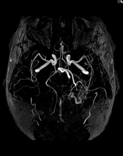
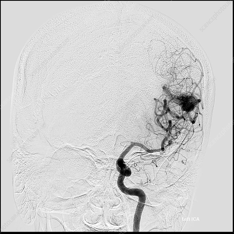
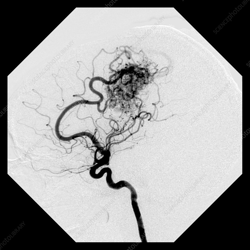
A brain AVM (arteriovenous malformation) is an abnormal tangle of blood vessels in which arteries connect directly to veins, bypassing the normal capillary network that should sit between them. Because capillaries are designed to slow blood flow, their absence means high-pressure arterial blood flows directly into the thin-walled veins.

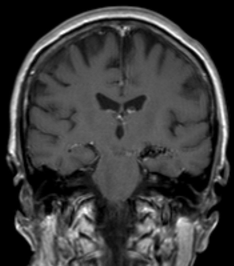
AVMs are usually present from birth and grow over time. Many AVMs never cause problems and are found only when imaging is done for another reason. Others can rupture and bleed into the brain, cause seizures, or produce progressive neurological symptoms depending on their location.

AVMs are uncommon — roughly 1 in 1,000 people have one — but they are one of the most common causes of hemorrhagic stroke in children and young adults.

Schematic illustration of a cerebral arteriovenous malformation showing a feeding artery, tangled nidus, and dilated draining vein
Symptoms

Symptoms and Warning Signs

Many AVMs cause no symptoms at all. When symptoms do appear, they vary depending on the AVM's size and location:

Seizures — often the first sign that an AVM is present
Headaches — sometimes severe, localized to one area
Weakness, numbness, or paralysis on one side of the body
Vision changes or loss of vision in one area
Difficulty speaking or understanding language
Confusion, memory problems, or personality changes
Sudden, severe headache — a sign of rupture, which is a 911 emergency
Time-of-flight MRA showing a brain AVM, rotated projection view
!
When to Call 911
A sudden, severe headache — particularly one that is unlike any headache you have had before — can be a sign that an AVM has ruptured. This is a life-threatening medical emergency. Call 911 immediately. Do not wait for symptoms to improve.
Treatment

Treatment Options

Not every AVM needs treatment. When treatment is recommended, the choice depends on the AVM's size, location, the symptoms it is causing, and the patient's overall health. Most treatment plans involve more than one approach.

EM
Endovascular Embolization

A catheter is threaded from the wrist or groin up to the brain, and a liquid embolic agent is injected to block the abnormal vessels. Embolization is often used as a first step before surgery or radiosurgery to reduce the size of the AVM and lower the risk of the definitive treatment. In select smaller AVMs, embolization alone may be curative.

SR
Surgical Removal

Open neurosurgery to physically remove the AVM. This is the most definitive treatment when the AVM is in a location that can be safely reached. Our team works closely with cerebrovascular neurosurgeons when surgical removal is the recommended approach.

RS
Stereotactic Radiosurgery

A non-invasive, focused radiation treatment (such as Gamma Knife or LINAC) that targets the AVM with high precision. The radiation causes the abnormal vessels to gradually close off over 1–3 years. Best suited for smaller, deeper AVMs that are difficult to reach with surgery or embolization.

Frontal cerebral angiogram from a left internal carotid artery injection showing a high-flow AVM in the left temporal lobe
Approach

A Combined Approach for Most AVMs

Most AVMs are not treated with a single approach. The typical path is:

  1. Diagnostic cerebral angiogram to map the AVM in detail
  2. Endovascular embolization to reduce the AVM's size and lower the risk of the next step
  3. Surgical removal or radiosurgery to eliminate what remains
  4. Long-term follow-up imaging to confirm the AVM has not recurred

Your neurovascular team will work with you to choose the combination that fits your specific AVM. The goal is always to eliminate the AVM's risk of bleeding while preserving as much brain function as possible.

T1-weighted contrast-enhanced MRI in the coronal plane, showing an AVM
Procedure

What Is AVM Embolization?

Embolization is a minimally invasive procedure performed in an angiography suite. A catheter is threaded through the arteries up to the AVM, where a special liquid or material is injected to block the abnormal vessels from the inside.

1
Catheter Access
A thin catheter is inserted through an artery in the wrist or groin and guided up to the blood vessels in the brain using real-time X-ray imaging.
2
Angiographic Mapping
Contrast dye is injected to create a detailed map of the AVM, identifying the feeding arteries, the nidus, and the draining veins.
3
Selective Catheterization
A microcatheter is navigated into the specific branches of the feeding arteries that supply the AVM nidus.
4
Embolization
A liquid embolic agent (such as Onyx or a similar material) is carefully injected through the microcatheter, filling the AVM nidus and blocking the abnormal vessels.
5
Confirmation & Recovery
Final angiographic images confirm that the targeted vessels are sealed. The catheter is removed and the patient is monitored before discharge.
Lateral cerebral angiogram showing the typical appearance of a high-flow arteriovenous malformation
Frequently Asked Questions

AVM questions, answered

Related Conditions

Need an AVM evaluation?

Our neurovascular team specializes in the diagnosis and treatment of brain AVMs. Contact us to schedule a consultation.